
Understanding the Benefits of Medical Thermography
Medical Thermography is a cutting-edge tool in the world of medical imaging. It offers a non-invasive way to assess health. This technique uses thermal imaging to detect heat patterns in the body.
Unlike traditional methods, it does not use radiation. This makes it a safer option for frequent monitoring. It is particularly useful for early detection of various conditions.
Thermal imaging can reveal inflammation, a sign of many chronic diseases. It is also effective in monitoring musculoskeletal injuries. This includes conditions like arthritis and fibromyalgia.
Medical Thermography provides a visual map of body temperature. This helps identify areas of concern. It is a valuable tool in sports medicine for injury detection and recovery monitoring.
The technology is advancing rapidly. Newer thermal cameras are more sensitive and accurate. This enhances its diagnostic capabilities.
Medical Thermography is gaining popularity in preventive healthcare. It allows for early intervention before symptoms appear. This makes it a cost-effective and accessible option for many.
What is Medical Thermography?
Medical Thermography is a revolutionary diagnostic tool. It employs infrared technology to visualize heat patterns on the skin’s surface. These patterns can indicate underlying health issues.
The technology is similar to that found in thermal cameras. It captures images that show variations in temperature. Warmer areas may indicate inflammation or increased blood flow.
This method is known for its non-invasive nature. It doesn’t require any physical contact. Patients find it comfortable and stress-free.
Medical Thermography has a broad range of applications. It detects unusual heat patterns linked to tumors. This application is particularly vital in breast health monitoring.
Beyond its imaging capabilities, the technology is groundbreaking. It offers insights into areas traditional methods may overlook. This innovation is pivotal in early disease detection.
One key feature is its radiation-free approach. This makes it suitable for regular screening without health risks. Patients appreciate its safe, consistent monitoring ability.
Some common uses include:
- Identifying inflammation and pain points
- Monitoring blood flow and circulation
- Detecting breast cancer at early stages
- Assessing musculoskeletal injuries
Advancements in this field continue to evolve. The refinement of thermal cameras has improved accuracy. This progress promises to expand its clinical utility.

The technology has piqued interest across medical disciplines. Its development and application continue to grow. As a result, its potential to revolutionize patient care increases. Thus, Medical Thermography remains a compelling innovation in diagnostic imaging.
The Science Behind Thermal Imaging
The foundation of thermal imaging lies in physics. All objects emit infrared radiation, which varies with temperature. This radiation is invisible to the naked eye.
Thermal imaging cameras convert this infrared data into clear images. They create visual representations of temperature differences. These images help to detect changes in the body’s heat patterns.
The process starts with the camera detecting infrared radiation. The camera’s sensors translate this data into an electronic signal. This signal forms a vivid thermographic image.
Medical Thermography uses these images for diagnostic purposes. The varying shades represent temperature variations. Hotter areas could indicate inflammation or excessive blood flow.
This technology offers real-time imaging possibilities. It provides immediate feedback about temperature changes in the body. This feature is invaluable for monitoring dynamic physiological processes.
Thermal imaging doesn’t require exposure to radiation or contrast agents. This makes it a safe, non-invasive option for many patients. As a result, it is used extensively in both clinical and non-clinical settings.
Applications of thermal imaging in medicine include:
- Early detection of breast cancer
- Monitoring of vascular disorders
- Assessment of muscular injuries
- Evaluation of chronic pain syndromes
The intricacies of thermal imaging extend beyond basic operation. It involves calibration and precision to ensure accuracy. The expertise of technicians and clinicians is crucial in interpreting results.

The science behind thermal imaging continues to advance. Innovations in sensor technology enhance resolution and sensitivity. These developments ensure that thermal imaging remains a vital tool in modern diagnostics. The field’s expanding scope promises new healthcare applications.
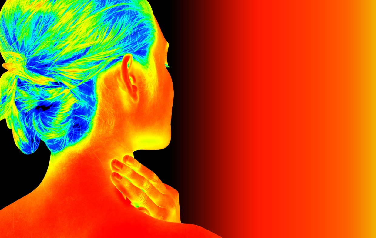
How Medical Thermography Works
Medical Thermography relies on sophisticated technology to map heat patterns. These patterns reveal insights into the body’s internal state. By detecting minute temperature changes, it aids in diagnosis.
The process begins with the patient standing or sitting in a controlled environment. This setting minimizes external temperature influences. A thermal camera captures infrared energy emitted from the body.
These cameras convert infrared radiation into temperature readings. This data translates into thermographic images. These images display variations in temperature with different colors.
Red or orange shades often indicate warmer areas. This could suggest inflammation, increased blood flow, or potential issues. Cooler areas appear in blue or green, hinting at reduced circulation or nerve issues.
The procedure requires no physical contact. Patients remain comfortable throughout. It is painless and poses no risk of radiation exposure.
The interpretation of thermographic images is key. Trained professionals analyze color patterns to identify anomalies. They correlate these patterns with physiological conditions.

by Logan Voss (https://unsplash.com/@loganvoss)
Applications where Medical Thermography excels include:
- Detection of breast abnormalities
- Monitoring of inflammatory processes
- Evaluation of post-surgery healing
- Assessment of chronic pain areas
The accuracy of this technology depends on the quality of the thermal camera. Skilled technicians are critical for meaningful analysis. Their expertise determines the reliability of the findings.
Medical Thermography evolves with advancements in imaging technology. New developments promise even greater precision. Such innovation expands its diagnostic potential across various medical fields.
Key Benefits of Medical Thermography
Medical Thermography offers several unique benefits. As a non-invasive technique, it opens doors to safer diagnostic possibilities. Its radiation-free nature makes it ideal for frequent monitoring.
One of the standout benefits is early detection. Thermography identifies subtle heat changes before symptoms appear. This can lead to timely intervention and prevention.
Thermal imaging serves as an excellent diagnostic supplement. It enhances other imaging techniques by providing additional information. This comprehensive view aids in accurate diagnosis.

by Logan Voss (https://unsplash.com/@loganvoss)
Key Advantages of Medical Thermography:
- Non-invasive, no direct body contact
- No exposure to harmful radiation
- Detects potential issues early
Thermography’s pain-free process is another merit. Patients remain at ease throughout the procedure. It suits those with anxiety over invasive methods.
The technology can evaluate multiple conditions effectively. It is valuable in assessing breast health and vascular issues. Chronic pain and inflammation also benefit from detailed thermal analysis.
Cost-effectiveness is an essential benefit. Compared to other medical imaging techniques, it is more affordable. This makes it accessible to a broader range of patients.
Thermography’s versatility extends beyond human health. It’s applicable in veterinary medicine as well. Animals can be examined without distress or sedation.
The technology provides a visual map of body temperature. This map facilitates pinpointing problem areas swiftly. It guides both diagnostics and treatment plans.
Applications in Various Fields:
- Early detection of tumors and inflammation
- Monitoring recovery in sports medicine
- Diagnosing circulatory disorders
Continual advancements in thermal cameras promise further benefits. These developments enhance resolution and accuracy. As technology improves, so does the scope of applications.
Patients and healthcare providers are recognizing these advantages. Thermography is becoming integral in preventive care strategies. Its growing popularity marks a shift toward non-invasive diagnostics.
Medical Thermography vs. Other Medical Imaging Techniques
Medical Thermography stands apart from traditional imaging methods. Unlike X-rays or CT scans, thermography uses no radiation. This absence of radiation ensures safety for frequent use.
Thermography detects heat variations in the body. It does not visualize internal structures, unlike MRI or ultrasound. Despite this, it provides valuable information through thermal data.

by Abdulai Sayni (https://unsplash.com/@abdulaisayni80)
Other imaging techniques focus on anatomical features. Thermography, however, highlights physiological changes. This difference can be critical for early disease detection.
Contrasting Imaging Features:
- X-ray/CT Scan: Uses radiation, shows bone and tissue structure.
- MRI/Ultrasound: No radiation, provides detailed internal images.
- Thermography: No radiation, captures heat patterns and blood flow.
Thermal imaging offers unique advantages in specific scenarios. It’s particularly effective for monitoring inflammatory processes. Other imaging methods may not detect these early functional changes.
For patients with allergies or contraindications to dyes, thermography is a viable option. It avoids the need for any injectable contrast agents. This reduces potential allergic reactions significantly.
In some cases, thermography enhances findings from other techniques. For instance, it can pinpoint areas needing further exploration with more detailed imaging. This approach results in a more comprehensive diagnostic picture.
It’s vital to recognize the complementary nature of thermography. While it provides unique insights, it often works best alongside other imaging techniques. Together, they can provide a holistic view of a patient’s health.
Medical Thermography complements existing imaging methods, offering a different perspective. Its ability to detect functional abnormalities before structural changes appear can be invaluable. Such an approach enhances early intervention strategies, ultimately benefiting patient outcomes.
Applications in Early Detection and Diagnosis
Medical Thermography has significant applications in early disease detection. Its ability to detect abnormal heat patterns is crucial. These patterns often indicate inflammation or disease processes.

by Margaret Kester (https://unsplash.com/@margaretkester)
Thermal imaging detects changes before symptoms appear. This early detection allows for timely intervention. Early interventions can dramatically improve health outcomes.
Key applications include:
- Breast Cancer: Identifies abnormal heat patterns before tumors grow.
- Inflammatory Conditions: Detects early signs of arthritis and similar diseases.
- Circulatory Disorders: Highlights poor blood flow areas early on.
Medical Thermography is valuable for detecting breast cancer. It can identify heat anomalies associated with tumor growth. Such early identification can significantly aid in early treatment planning.
Benefits of Early Detection with Thermography
- Non-invasive Monitoring: No need for invasive procedures or radiation exposure.
- Early Intervention: Allows for treatment before disease progression.
- Enhanced Surveillance: Facilitates continuous health monitoring over time.
Thermography is beneficial in monitoring inflammatory diseases. Conditions like rheumatoid arthritis show heat changes in early stages. Catching these early signs can prevent further joint damage.
In vascular health, thermal imaging identifies circulation issues. Detecting areas of poor circulation helps prevent serious complications. For example, thermal anomalies can signal potential ulceration in diabetes.
Sports medicine also uses thermography for early injury detection. Before an injury becomes severe, changes in thermal patterns can be detected. This ensures proper management and swift recovery.
Using thermography for diagnosis extends beyond identifying existing conditions. It can assess risks and predict future health issues. Monitoring subtle temperature changes offers predictive insights.
Medical Thermography continues to play a pivotal role in preventive health care. With its ability to detect changes early, it supports proactive health strategies. By integrating thermography into routine checks, healthcare providers can manage potential health issues more effectively.
Overall, the early detection capabilities of thermography enhance patient management. They allow healthcare providers to act before conditions worsen. Early diagnosis remains a cornerstone of preventive medicine, and Medical Thermography proves to be a powerful tool in this domain.
Breast Thermography: A Closer Look
Breast Thermography offers a unique approach to breast health monitoring. It serves as a supplemental tool to traditional imaging methods like mammography. Its strength lies in its ability to detect physiological changes early.

by Logan Voss (https://unsplash.com/@loganvoss)
This method involves using infrared cameras. These cameras capture heat patterns of breast tissue. Tumors often disrupt normal thermal patterns, making them detectable.
Key advantages of breast thermography include:
- Non-invasive: No direct contact with the body is necessary.
- Radiation-free: Unlike mammograms, no radiation exposure occurs.
- Early Detection: Identifies potential issues before they become palpable.
Despite its benefits, it’s crucial not to rely solely on thermography. It is best used alongside traditional diagnostic methods. Combining these tools enhances the detection of any anomalies.
Breast Thermography is particularly useful for dense breast tissue. Dense tissue makes mammogram readings more challenging. Thermal imaging provides another layer of information, reducing uncertainty.
This technique suits women at higher risk of breast cancer. For example, those with a family history of breast cancer can benefit from frequent thermographic assessments. It provides an ongoing evaluation without the drawbacks of repeated radiation exposure.
To maximize accuracy, practitioners should have expertise in interpreting thermal images. Accurate interpretation is vital for identifying suspicious thermal patterns. Experienced practitioners can help ensure the best use of thermal imaging data.
Overall, breast thermography is gaining traction. As technology advances, its role in breast health management may expand further. By integrating thermography into regular breast health assessments, women can be more proactive about their well-being. It’s an important development in the fight against breast cancer and a step forward in promoting women’s health.
Use in Musculoskeletal and Pain Disorders
Medical Thermography is extremely useful in evaluating musculoskeletal disorders. It helps identify inflammation in joints and tissues, which is essential in conditions like arthritis.

by Google DeepMind (https://unsplash.com/@googledeepmind)
The technique provides a clear visual of heat distribution. Abnormal heat patterns may indicate inflammation or injury. This is particularly important in conditions like fibromyalgia where pain is widespread.
Using thermal imaging in musculoskeletal disorders offers several benefits:
- Non-invasive: A safe method without needing physical contact.
- Real-time monitoring: Allows for ongoing tracking of inflammation levels.
- Objective analysis: Visual evidence of temperature changes in affected areas.
Understanding pain disorders requires more than just patient history. Thermography provides an additional layer of data that enhances diagnosis and treatment strategies.
These images can reveal subtle temperature differences caused by blood flow changes. This is important for assessing soft tissue injuries, including tendinitis and bursitis. By visualizing these changes, practitioners can make more informed decisions about interventions.
One of the most significant advantages is in sports medicine. Athletes often need to monitor recovery from injuries closely. Thermography helps in tracking progress without imposing additional stress on the affected area.
In chronic pain management, thermography assists in evaluating the effectiveness of treatments. By observing changes in heat patterns over time, healthcare providers can adjust therapies to better suit individual needs.
Adopting thermographic techniques in the assessment and monitoring of musculoskeletal disorders improves patient care. It allows for a comprehensive understanding of the underlying causes of pain. As a result, both the detection and management of these conditions are enhanced. This contributes to better patient outcomes and more personalized treatment plans.
Vascular and Circulatory Assessments
Medical Thermography offers unique insights into vascular and circulatory health. It does so by highlighting heat patterns linked to blood flow.

by Logan Voss (https://unsplash.com/@loganvoss)
In vascular assessments, thermography identifies areas of poor circulation. These areas often display cooler regions due to reduced blood flow. This non-invasive approach is particularly beneficial for patients with vascular disorders who need regular monitoring.
Thermal imaging is adept at detecting varicose veins. The heat variations signify compromised veins, aiding timely diagnosis. This is a crucial step in preventing severe complications like ulcers or thrombosis.
Thermography’s role is expanding in detecting peripheral artery disease (PAD). By identifying thermal anomalies, it helps pinpoint areas with restricted blood flow. Early detection is vital as PAD can lead to severe complications, such as heart attacks or strokes.
The advantages of using thermography for vascular assessments include:
- Radiation-free: Safeguards patients from harmful exposure.
- Pain-free: Comfortable with no need for sedation or injections.
- Comprehensive: Provides a broad view of circulatory status without invasive procedures.
For people with diabetes, monitoring circulation is crucial. Thermography detects early signs of reduced blood flow in extremities. This early warning is vital for preventing foot ulcers and other complications.
Thermography complements other diagnostic tools in vascular care. When integrated into regular check-ups, it enhances disease management. This leads to timely interventions, potentially reducing severe outcomes. Overall, the incorporation of thermal imaging in vascular assessments enhances clinical practice by providing real-time, insightful data.
Role in Sports Medicine and Injury Recovery
Medical Thermography plays a pivotal role in sports medicine. Athletes benefit from its ability to detect injuries early. This is essential for both professional and amateur athletes.

by Alina Belogolova (https://unsplash.com/@alinabelogolova)
Thermal imaging identifies inflammation indicative of injuries. Anomalies in heat patterns can point out sprains, strains, and muscle tears. These insights allow for quicker intervention and treatment.
Post-injury, thermography assists in monitoring recovery. Doctors can track changes in heat distribution over time. This method offers a clear view of healing progress.
The technique is particularly useful in customizing rehabilitation strategies. By visualizing areas of concern, practitioners tailor therapy plans. This personalized approach enhances recovery outcomes.
Medical Thermography’s benefits in sports medicine include:
- Non-invasive monitoring: No need for physical probes or tools.
- Frequent assessments: Safe for regular use without radiation risk.
- Detailed visualization: Offers clear images of soft tissue status.
By focusing on areas of altered blood flow, thermography guides athletes back to peak performance. This imaging is crucial in optimizing training regimens. Detecting minor injuries before they worsen minimizes downtime.
Incorporating thermography into sports practice optimizes care. Athletes return to competition more confidently and swiftly. Overall, it’s an invaluable resource in both preventing and managing sports-related injuries.
Monitoring Chronic Conditions and Treatment Progress
Medical Thermography is a powerful tool for monitoring chronic conditions. It maps heat distribution and blood flow changes. This offers valuable insights for ongoing healthcare management.

by Roxy Aln (https://unsplash.com/@roxy_aln)
People with chronic conditions can benefit from this technology. Thermography detects subtle changes that indicate disease progression. This allows for timely interventions and adjustments in treatment plans.
Thermography is non-invasive and safe for repeated use. This makes it ideal for long-term monitoring. It avoids the risks associated with radiation exposure from other imaging techniques.
In chronic pain management, thermal imaging highlights inflammation and other heat anomalies. These indicators help healthcare providers tailor treatment approaches. Accurate monitoring aids in assessing treatment effectiveness and patient response.
Medical Thermography offers several benefits for chronic condition monitoring:
- Regular assessments: Safe for frequent imaging sessions.
- Progress tracking: Monitors therapeutic impact over time.
- Non-invasive approach: No need for invasive probes or contrast dyes.
Practitioners can adjust therapies based on thermographic findings. This proactive approach can improve patient outcomes. By visualizing treatment responses, doctors make data-driven decisions.
For chronic disease patients, this imaging can be transformative. It allows for a detailed look at physiological changes, which might be missed using traditional methods. Early detection of flare-ups can lead to better management strategies.
Veterinary and Non-Human Applications
Medical Thermography isn’t just beneficial for humans; it’s also a vital tool in veterinary medicine. Animals can’t easily communicate discomfort or pain. Thermal imaging helps veterinarians detect issues that might otherwise go unnoticed.
Thermography provides a visual representation of heat patterns on an animal’s body. This can identify areas of inflammation, injury, or disease. The technique is non-invasive and quick, minimizing stress for animals during examination.
The use of thermal imaging in veterinary care includes:
- Detecting joint inflammation in horses.
- Monitoring skin conditions in cats and dogs.
- Assessing overall health in livestock.
Beyond pets, thermography assists zookeepers and wildlife researchers. It aids in the health assessment of exotic and endangered species. Observing thermal changes helps in making informed decisions for animal care and welfare.
With ongoing advancements, thermography continues to expand its applications. As sensitivity and accuracy improve, this tool becomes even more crucial for ensuring the well-being of animals.
Safety, Comfort, and Accessibility
Medical Thermography offers an exceptionally safe imaging option. Unlike X-rays or CT scans, it uses no ionizing radiation. This absence of radiation reduces the risk associated with repeated scans, making it a safer choice for long-term monitoring.

by Shanjir H (https://unsplash.com/@shanjir)
Comfort during the procedure is another advantage. Patients experience no physical contact, as the thermal camera captures heat emitted from the body from a distance. This feature is particularly beneficial for individuals sensitive to touch or pressure.
Thermography’s accessibility further enhances its appeal:
- Non-invasive, with no need for injections or dyes.
- Quick procedure, often completed in less than 30 minutes.
- Suitable for all ages, from children to the elderly.
This combination of safety, comfort, and accessibility ensures Thermography is well-suited to a wide array of patients. From chronic illness monitoring to pain management, it serves diverse needs. Additionally, the process is efficient and straightforward, fostering a positive experience for patients.
As awareness grows, more clinics and facilities are incorporating this technology. This broader availability means more individuals can benefit from its non-invasive insights into health.
Limitations and Considerations
While Medical Thermography offers many benefits, it’s important to acknowledge its limitations. One primary concern is the dependency on environmental conditions. A controlled environment is crucial for accurate thermal readings, as temperature fluctuations can affect the results.

by Adi-DE (https://unsplash.com/@adasko73)
Another consideration is the requirement for specialized expertise. Skilled interpretation is vital to avoid false positives or negatives. Without a qualified professional, the analysis might miss important thermal anomalies.
Despite these challenges, Thermography is a valuable tool when used alongside traditional imaging techniques. It provides complementary data rather than a standalone diagnosis. Understanding its role within a broader diagnostic context is essential for maximizing its benefits.
Patients should also be informed about the scope and limitations of Thermal Imaging. This knowledge helps set realistic expectations and reduces misunderstandings. Comprehensive consultation is crucial for an informed decision-making process.
Awareness of insurance coverage is another practical aspect. While some insurers recognize Thermography, coverage varies significantly. Patients should verify their policy details before proceeding with the test to avoid unexpected costs. Exploring insurance options can help determine the affordability and accessibility of this imaging method.
Technological Advancements in Medical Thermography
Recent years have seen significant advancements in Medical Thermography technology. The development of more sensitive thermal cameras has greatly enhanced image resolution. These improvements make it easier to detect subtle heat patterns crucial for accurate diagnostics.

by Projector1 (https://unsplash.com/@projector1)
One of the major breakthroughs is the integration of artificial intelligence in image analysis. AI technologies assist in interpreting complex thermal data swiftly and accurately. This reduces human error and leads to more reliable diagnostic outcomes.
In addition to AI, real-time monitoring capabilities have also evolved. New systems allow continuous observation of thermal changes, offering dynamic insights into a patient’s condition. This is particularly beneficial in tracking recovery or response to treatments.
Cost reduction has made Thermal Imaging more accessible. Advances in manufacturing processes have led to lower production costs for cameras. Consequently, more healthcare providers can afford to incorporate this technology into their practice.
Some key advancements include:
- High-resolution thermal sensors.
- Enhanced AI-based diagnostic tools.
- Real-time monitoring systems.
- Streamlined and portable designs.
These developments represent a promising future for Medical Thermography. As technology continues to evolve, so will the applications and effectiveness of this invaluable tool in medical imaging. Embracing these innovations will likely expand its role in preventive and diagnostic healthcare, offering broader applications and benefits.
Integrating Thermography into Preventive Healthcare
Medical Thermography is not just for diagnosis; it’s pivotal in preventive healthcare. By detecting heat patterns, it identifies potential issues before symptoms emerge. This early detection can lead to proactive treatments, reducing the risk of severe health conditions.

by Brian Wangenheim (https://unsplash.com/@brianwangenheim)
Thermography’s non-invasive nature makes it ideal for regular health checks. It eliminates the risks associated with radiation exposure from other imaging methods. This feature is particularly beneficial for those requiring frequent monitoring.
Integrating thermal imaging in routine check-ups helps maintain better health outcomes. Patients and healthcare providers can monitor inflammation and other anomalies continuously. It supports timely interventions, which play a vital role in preventive strategies.
Moreover, thermography encourages a holistic approach to wellness. By identifying systemic imbalances, it aids in tailoring personalized health plans. This integration aligns with modern healthcare trends focusing on long-term well-being rather than just treating symptoms.
Key applications in preventive care include:
- Monitoring inflammation to prevent chronic diseases.
- Tracking vascular health and circulation issues.
- Routine checks in wellness programs.
- Early detection of tissue anomalies.
As healthcare systems strive to prevent diseases rather than merely treat them, Medical Thermography offers an effective tool for achieving this goal. Embracing this technology may lead to healthier populations and lower healthcare costs long-term.
Patient Experience: What to Expect During a Thermography Session
Undergoing a Medical Thermography session is straightforward and stress-free. The process begins with a brief consultation to discuss health history and any concerns. This step helps tailor the procedure to the patient’s specific needs.

by FilterGrade (https://unsplash.com/@filtergrade)
Patients are asked to acclimatize to room temperature, which often takes about 15 minutes. This ensures accuracy by eliminating any external temperature influences. It’s a non-contact process, meaning there is no physical touch involved with the imaging equipment.
During the actual imaging, patients stand or sit in front of an infrared camera. The procedure is quick, typically lasting about 10 to 15 minutes. There are no uncomfortable positions, making it ideal for people of all ages and conditions.
After the session, patients receive a report detailing any heat patterns or anomalies. This report aids in understanding potential health issues and guiding further medical consultations. Understanding what to expect demystifies the process and enhances patient comfort and confidence.
Key steps in a session include:
- Initial consultation
- Room acclimation
- Non-contact imaging
- Receiving a detailed report
Choosing a Qualified Thermography Provider
Selecting a reliable thermography provider is crucial for accurate assessments. Providers should possess appropriate certifications and experience in thermal imaging. These credentials ensure they follow established protocols and interpret results correctly.

by Maria Luisa del Carmen Romero Centeno (https://unsplash.com/@luisaromero)
In addition to credentials, consider the provider’s equipment. Modern, sensitive cameras significantly enhance accuracy. Advanced technology enables detailed, reliable results that older equipment may not provide.
Customer reviews and feedback can offer valuable insights. Look for providers with positive testimonials regarding both their service and expertise. These reviews often reflect the provider’s ability to deliver a professional and comforting experience.
Lastly, consider the provider’s willingness to discuss findings and implications. A qualified provider should explain the results clearly. They should be open to questions and guidance on further steps if necessary.
Here are points to consider when choosing a provider:
- Certifications and experience
- Modern equipment
- Positive customer reviews
- Clarity in result discussion
Insurance, Cost, and Accessibility
The cost of medical thermography varies by location and provider. Generally, it is considered a cost-effective diagnostic tool. Despite its affordability, coverage by insurance providers may be inconsistent.

by Marek Studzinski (https://unsplash.com/@jccards)
Some insurance companies recognize the benefits of thermography and include it in their plans. It’s important to verify with your insurance provider before scheduling a procedure. This can prevent unexpected out-of-pocket expenses.
For those without insurance coverage, price transparency is key. Many providers offer detailed pricing information upfront. This allows patients to plan financially and decide if thermography suits their budget.
Thermography’s non-invasive nature makes it accessible to a broad range of patients. Its accessibility ensures that even those who can’t undergo traditional imaging have an option. This makes it an ideal tool for diverse healthcare needs.
Consider these aspects regarding thermography’s cost and accessibility:
- Insurance coverage variability
- Importance of upfront pricing
- Non-invasiveness enables accessibility
Frequently Asked Questions about Medical Thermography
Is Medical Thermography Safe?
Yes, medical thermography is completely safe. It is a non-invasive procedure that emits no radiation. This makes it suitable for frequent monitoring without any harmful effects.
How Does Medical Thermography Differ from Other Imaging?
Unlike traditional imaging, thermography detects temperature variations. This helps in identifying inflammation and abnormal blood flow. Thus, it’s a unique tool in the field of medical imaging.
What Conditions Can Be Monitored?
Medical thermography can monitor various conditions, including musculoskeletal disorders and breast health issues. It also assists in tracking treatment effectiveness over time. Additionally, it is useful for sports injuries and chronic pain management.
Consider the following benefits:
- Radiation-free
- Non-invasive
- Detects temperature and blood flow changes
How Should I Prepare for a Session?
Preparation for a thermography session is simple. Avoid any skin creams, lotions, or deodorants before the procedure. Ensure the skin is clear to get the most accurate results possible. Additionally, avoid exercise before the session as it can affect temperature readings.
The Future of Medical Thermography
The future of medical thermography looks promising with ongoing technological advancements. Emerging technologies are making thermal imaging more precise and accessible. This progress is expected to enhance diagnostics significantly.
Improvements in thermal camera sensitivity enable detection of subtler temperature variations. This helps identify conditions even earlier, improving patient outcomes. As technology evolves, thermography’s diagnostic accuracy will continue to improve.
The integration of artificial intelligence (AI) is another exciting development. AI algorithms analyze thermal images more quickly and effectively. This leads to faster diagnosis and can provide more personalized treatment options.
Consider these future possibilities:
- Enhanced image resolution
- Faster image processing
- Greater access and reduced costs

by Wolfgang Hasselmann (https://unsplash.com/@wolfgang_hasselmann)
Growing interest in preventive healthcare will likely increase thermography’s applications. As more people seek non-invasive diagnostic options, its popularity is set to rise. This shift will further establish medical thermography as a staple in modern healthcare. The potential to revolutionize diagnostic processes is substantial, aiming for a healthier future.
Conclusion: Embracing the Benefits of Medical Thermography
Medical thermography offers a remarkable glimpse into the body’s health through thermal patterns. This non-invasive tool helps in early detection and supports preventive healthcare.
Compared to traditional imaging methods, thermography provides a safer and more comfortable experience. Without radiation exposure, frequent monitoring becomes risk-free, making it ideal for continuous health assessments.
As technology advances, medical thermography will grow in its applications and capabilities. Embracing this innovative approach opens new avenues in diagnostics and patient care. By integrating thermography into standard medical practices, healthcare providers can enhance their diagnostic toolkit, leading to earlier interventions and improved health outcomes. The potential of medical thermography in shaping the future of healthcare is immense.


Leave a Reply
Your email is safe with us.